
Lewisburg Animal Hospital is proud to offer general digital radiology, digital dental radiology, and ultrasonography.
Lewisburg Animal Hospital is proud to offer general digital radiology, digital dental radiology, and ultrasonography.
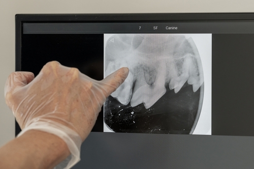
Dental radiography is a vital component of our dental procedures. Many dental issues cannot be determined simply by examining the crown of the tooth. Dental X-rays allow us to examine below the gumline by visualizing the roots of the teeth. We can identify abscesses or other dental diseases that may need treatment.

Radiographs, or x-rays, are commonly used to diagnose orthopedic or soft tissue diseases such as fractured bones, internal masses, bladder stones, spinal disorders, heart/lung disease, enlarged organs, pregnancy diagnosis, and diagnosis of foreign bodies in the gastrointestinal tract. In addition, we can submit radiographs to board-certified veterinary radiologists for review. This is like having a board-certified radiologist on staff and is great for second opinions and more complex cases.
We can x-ray your dog’s hips and/or elbows to screen for dysplasia. If desired, the X-rays can be sent to the Orthopedic Foundation of America (OFA) where board-certified radiologists will evaluate and grade your dog’s hips and/or elbows for OFA certification. To capture these X-rays, the patient is generally sedated or anesthetized to obtain proper positioning.
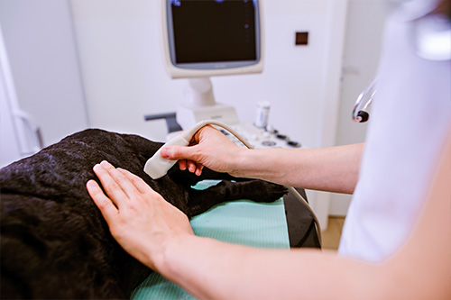
Ultrasonography (also called ultrasound or sonography) is a noninvasive, pain-free procedure that uses sound waves to examine a pet’s internal organs and other structures inside the body. It can be used to evaluate the animal’s heart, kidneys, liver, gallbladder, and bladder; to detect fluid, cysts, tumors, or abscesses; and to confirm pregnancy or monitor an ongoing pregnancy.
We may use this imaging technique in conjunction with radiography (x-rays) and other diagnostic methods to ensure a proper diagnosis. Interpretation of ultrasound images requires great skill on the part of the clinician.
The ultrasonographer applies gel to the surface of the body and then methodically moves a transducer (a small handheld tool) across the skin to record images of the area of interest. The gel helps the transducer slide more easily and creates a more accurate visual image.
The transducer emits ultrasonic sound waves, which are directed into the body toward the structures to be examined. The waves create echoes of varying degrees depending on the density of the tissue and the amount of fluid present. Those waves create detailed images of the structures, which are shown on a monitor and recorded for evaluation.
Ultrasound does not involve radiation, has no known side effects, and doesn’t typically require pets to be sedated or anesthetized. The hair in the area to be examined usually needs to be shaved so the ultrasonographer can obtain the best result.
In more difficult cases, we may have a specialist ultrasound your pet. This can be done by a mobile ultrasonographer or at a referral practice. If we feel this is the best option for your pet, we will discuss this with you.
If you have any questions about our ultrasonography service or what to expect during your pet’s procedure, please don’t hesitate to ask.
1113 B East Commerce Street
Lewisburg, TN , 37091
Phone: (931) 359-5945
Monday: 7:45am – 5:30pm
Tuesday: 7:45am – 5:30pm
Wednesday: 7:45am – 5:30pm
Thursday: 7:45am – 5:30pm
Friday: 7:45am – 5:30pm
Saturday: 7:45am – 4:00pm
Sunday: Closed